In Vivo Imaging Core Facility
Who We Are
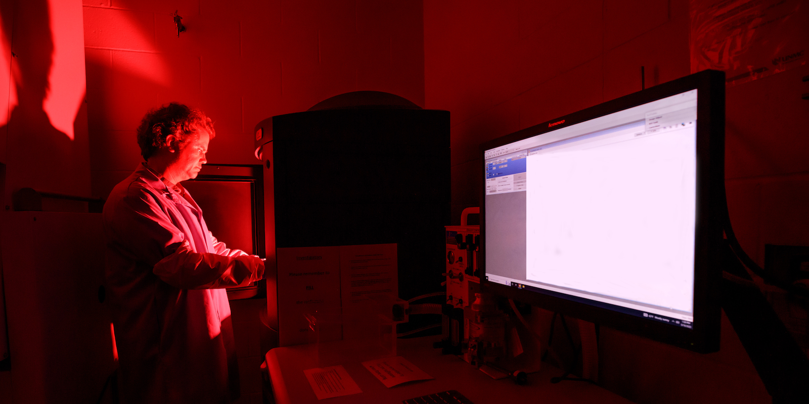
The In Vivo Imaging core facility is comprised of a sensitive CCD camera, a dark imaging chamber to minimize incident light, and specialized software to quantify and analyze results. The system features high-sensitivity in vivo imaging of fluorescence and bioluminescence, high throughput (5 mice) with 23 centimeter field of view, high resolution (to 20 microns) with 3.9 centimeter field of view, twenty eight high efficiency filters spanning 430 – 850 nanometer. An optical switch in the fluorescence illumination path allows reflection-mode or transmission-mode illumination. The system uses 3D diffuse tomographic reconstruction for both fluorescence and bioluminescence and has the ability to import and automatically co-register CT or MRI images yielding a functional and anatomical context for your scientific data