Advanced Microscopy Core Facility
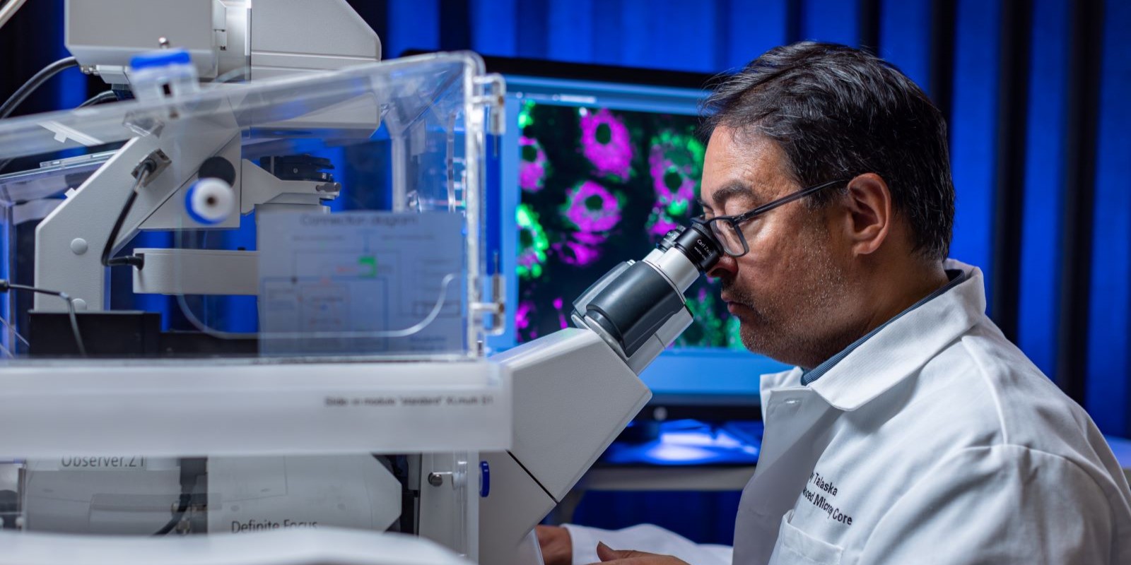
The Advanced Microscopy Core Facility at UNMC, a shared research resource supported by the Fred & Pamela Buffett Cancer Center and the NE-INBRE program, provides access to a diverse array of biomedical imaging modalities.
These tools are capable of capturing complex biological phenomena at single molecule (nanometer, super resolution), sub-cellular and cellular resolution (micron, confocal and whole slide scanning), and tissue/small organ levels (millimeter, light sheet). Instruction in the use of, and trained user access to imaging (super resolution, microscopic, mesoscopic) and analysis (HALO, IMARIS) instrumentation are available to internal and external researchers on a fee-for-use basis. With prior approval from the Director, the facility will also collect and/or process images for investigators on a fee-for-service basis.
The AMCF is located on the first floor of the Durham Research Center (DRC I, Rooms 1056, 1063, 1036, 1053). Requests for training and instrumentation access may be submitted by emailing or calling 402-559-6659.
Overview of Instruments and Services
The AMCF offers several instruments and services to support your research needs, including those listed below. To discuss individual imaging needs, contact the core director.
| Please note this list is not exhaustive. To explore the full range of our instruments and services, please visit our Instruments and Services page. | |
| Equipment/Service | Description |
|---|---|
| Zeiss Elyra PS.1 Superresolution Microscope | Single Molecule Localization Microscopy (PALM, STORM), Structured Illumination Microscopy (SIM) |
| Zeiss LSM 800 w/ Airyscan for High Resolution Imaging | Multi-Channel Imaging in fixed or live cells (incubated stages) |
| Zeiss 710 Confocal Laser Scanning Microscope | Dynamic Imaging: Time Series, FRAP, FRET, Spectral Imaging, 3D/Volumetric, High-Content Imaging |
| Zeiss Cell Discoverer 7 high Content Plate Reader | Multi-well, plastic plate imaging, Drug dispensing port |
| Zeiss Axioscan 7 Whole Slide Imaging System | 20x, 40x TL and FL Whole Slide Digitization |
| UltraMicroscope II Light Sheet Fluorescence Microscope | Mesoscopic, 3D/Volumetric Imaging of Small Organs, Tissues |
| X Clarity Automated Tissue Clearing System | Optical clearing for 3D/Volumetric Imaging |
| Other Services: |
Data Analysis Workstations – HALO, IMARIS, Zeiss Zen, QuPath Consultation – design, collection, analysis Training – hands-on across imaging modalities Education – in-person and online researcher resources Independent and Assisted Imaging and Analysis options |
Acknowledging Core Usage
Services and equipment in the AMCF are provided with support from multiple funding agencies and MUST be appropriately cited for sustained operation of these shared resources.
Researchers may now use a simplified core acknowledgement statement referencing our Research Resource ID (RRID).
“We acknowledge use of the University of Nebraska Medical Center - UNMC Advanced Microscopy Core Facility, RRID:SCR_022467, P20 GM103427 (NIGMS, NE-INBRE), P30 GM106397 (NIGMS, NCS), P20GM130447 (NIGMS, CoNDA), P30 CA036727 (NCI, Buffett Cancer Center), S10RR02730 (NIH), S10OD030486 (NIH), Nebraska Research Initiative, UNMC Vice Chancellor for Research Office.”
James R. Talaska, B.S., Camille Hennerberg, B.S., Heather Jensen-Smith, Ph.D.
AMCF users are obligated to fully acknowledge the facility and its funding sources in formal publications and presentations containing any data generated with support from the facility (instrumentation and/or staff).
Check out our Policies page for more information about acknowledgement statements, authorship guidelines, and usage of core instruments and services.