Mariano Uberti, PhD
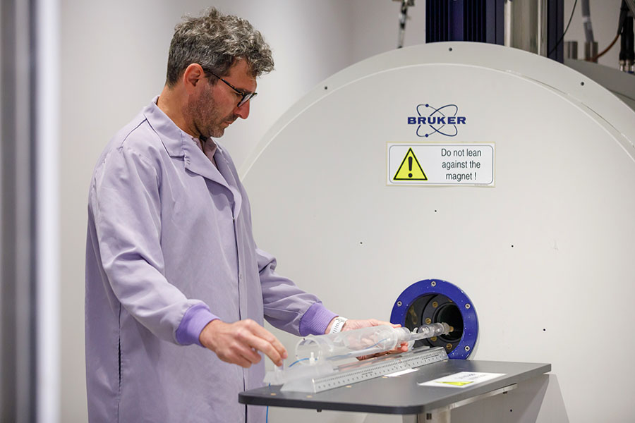
Mariano Uberti, PhD, prepares an experiment in the small animal MRI machine, a Bruker 7T scanner.
The scientific interests of Mariano Uberti, PhD, lie at the intersection of bioimaging technology development and its applications in biomedical research. His work integrates engineering and computer science tools to advance magnetic resonance imaging and spectroscopy methods for studying diseases, monitoring treatments, and aiding drug development.
His projects span from developing novel MRI methods to creating tools for streamlined and accurate image and signal data processing and visualization, as well as applying state-of-the-art in vivo MR modalities such as proton spectroscopy and chemical exchange saturation transfer MRI in both preclinical research with animal models and clinical research with human subjects.
Through these efforts, Dr. Uberti aims to push the boundaries of bioimaging technology and its applications, contributing to significant advancements in basic, translational, and clinical biomedical research.
Ongoing Projects
- Cancer Research: Studying the underlying glutamine metabolism of amide proton transfer MRI signals in glioblastoma.
- Clinical Research: Monitoring in vivo neurogenesis in encephalitis.
- Basic Research: Investigating brain metabolic imaging biomarkers of HIV-1 infection during antiretroviral therapy.
- Brain Molecular Connectivity: Using MRI and artificial intelligence technology to map brain molecular connectivity.
Tool Development
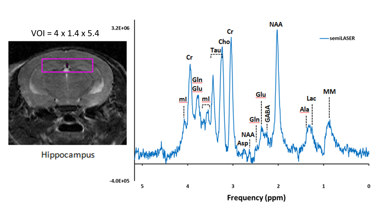
- Single-Voxel 1H-MRS semiLASER: Implementing and optimizing this method in a Bruker 7T scanner for more accurate and sensitive assessment of brain metabolites in small animals.
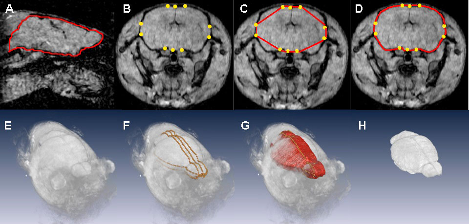
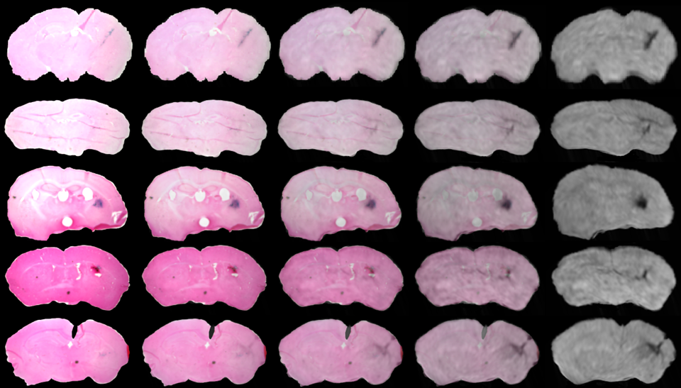
- Image Processing Software: Developing tools for murine brain MRI data, including semi-automatic brain extraction and co-registration of murine brain histological slices and MRI brain volumes using both linear and non-linear registration methods others.
- MRS Signal Processing Software: Creating automated and batch-capable processing and fitting routines, including quantitative assessment of different processing methods, basic statistical analysis, and interfacing with an Animal Tracking System database.
- Advanced 1H MRS Methods: Developing state-of-the-art 1H spectroscopy techniques in a 3T scanner for clinical research.