Neuroradiology
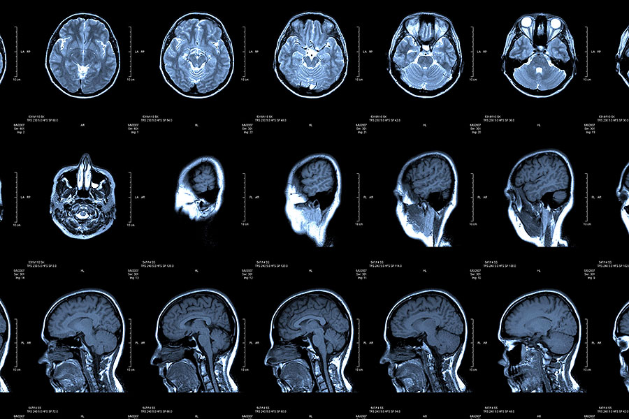
Our neuroradiologists specialize in diagnostic imaging techniques of the brain, the spine and the head and neck regions utilizing computed tomography and magnetic resonance imaging. Residents rotating through our division may experience:
- Myelography and lumbar puncture procedures performed with fluoroscopy.
- State-of-the-art imaging techniques implemented with CT and MR imaging that include perfusion, diffusion and spectroscopy.
- Noninvasive MR angiography, MR venography, CT angiography and CT venography imaging exams.
- Vessel wall imaging performed to evaluate complex cerebral vasculature cases to evaluate for vasculitis.
- Head and neck specialized imaging including orbit, ear, sinus, face, skull base, cranial nerve, soft tissue neck, thyroid and parathyroid examinations.

Matthew White, MD, Neuroradiology Division Chief
Dr. White is a prodigious scholar and expert in the field of functional MRI. He has been instrumental in the implementation of new neuro MR protocols at UNMC and has more than 25 years of experience in neuroradiology.
Brain tumors often distort the brain making it impossible to determine exactly where functional brain tissue lies on anatomical images. Our neuroradiology division has established a program to perform advanced brain tumor mapping in order to map gray matter function, the intricacies of white matter pathways and how these relate to brain tumors or other masses.
Neuroradiologists perform functional MRI utilizing numerous paradigms to evaluate language, auditory, vision, skin sensory and motor functions. To obtain a comprehensive understanding of language, paradigms are performed utilizing verb generation, word generation and/or sentence completion. Passive listening is utilized to test auditory activation. Motor functional analysis may involve finger tapping, foot tapping, and lip motion. Data is captured and analyzed in real time which allows neuroradiologists to tell if a certain activation paradigm is working or not.
Diffusion tensor imaging is based on the differential motion of water parallel to neural pathways in the brain versus perpendicular to neural pathways. Utilizing advanced computer analysis and an understanding of the neuroanatomy the white matter tracts can be mapped out. Diffusion tensor imaging white matter analysis can help demonstrate white matter tracts that are deviated or displaced by a tumor versus invaded or destroyed. Visualization of the deviation/displacement of the white matter neural pathways by a tumor can be determined in 3D space of the brain and how this relates to the brain tumor.
To provide as accurate of a “virtual brain lesion biopsy” as possible, our neuroradiology division has a long history of utilizing multiple advanced imaging techniques to determine what the underlying cause of brain lesions are. Magnetic resonance spectroscopy, brain perfusion analysis and diffusion weighted imaging analysis are available to analyze brain lesions. These techniques often help neuroradiologists to determine the likelihood of a brain lesion being a tumor.