Echocardiography Imaging
The echocardiography imaging facility consists of:
- Acuson 512C echocardiograph and associated probes (3-5 MHz and 15 MHz linear array).
- Visual Sonics Vevo 3100.
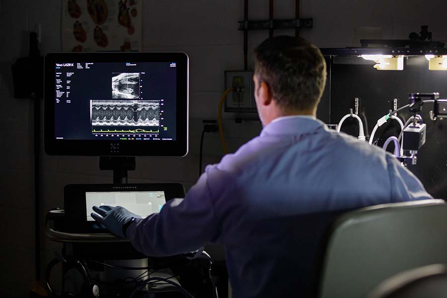
- Vevo 3100 with the LAZR-X.
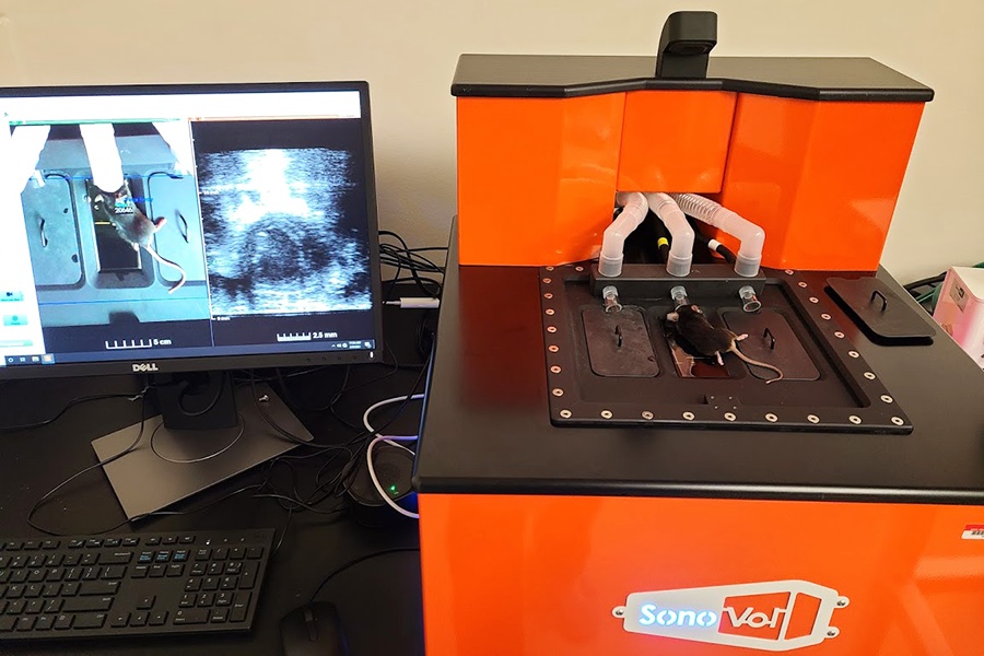
- Perkin Elmer Vega.
Each Vevo machine has probes ranging from 15 MHz to 55 MHz. The Vevo systems have injector rails for echo-guided delivery into rodents.
Cardiovascular imaging ranges from simple 2D and M-mode indices of cardiac function (EF, FS, dimensions, volumes, wall thickness, etc) to assessment of diastolic function (E/A) and pulsed Doppler flow, 4D imaging, and speckle tracking using Vevo Strain.
We also offer longitudinal imaging of tumors with available 3D volumetric analysis. Photoacoustic imaging with the LAZR-X uses a laser in the ranges of 680 nm - 970 nm or 1200 nm – 2000 nm to create a thermoelastic change in chromogens which generates a mechanical signal picked up by ultrasound. This can be used to detect changes in oxygen saturation levels of hemoglobin or other chromogens (ICG, IR-800, etc). Changes in flow can be monitored using non-linear contrast imaging and microbubbles.
The Vega system brings high throughput imaging to the core along with shear wave elastography and acoustic angiography.
Fees
The core operates on a fee-for-service basis. Charges range from $60 to $100 per hour, dependent on technician involvement. Analysis can be done by investigators or by the core director as an additional charge.
Director
Bryan T. Hackfort, PhD
Assistant Professor, UNMC Department of Cellular and Integrative Physiology
Director, three research cores
