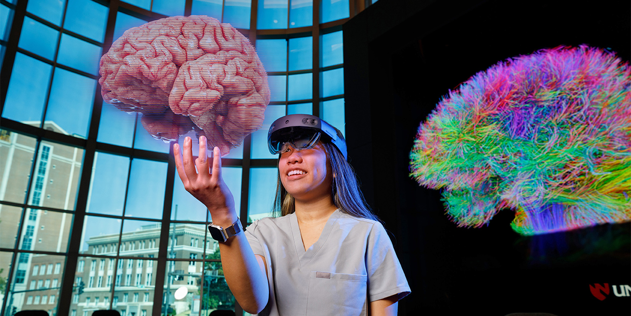
UNMC formed the iEXCEL program to reach a new level in 3D, virtual reality and augmented reality medical imagery, allowing health professions students to learn their skills in new, engaging ways.
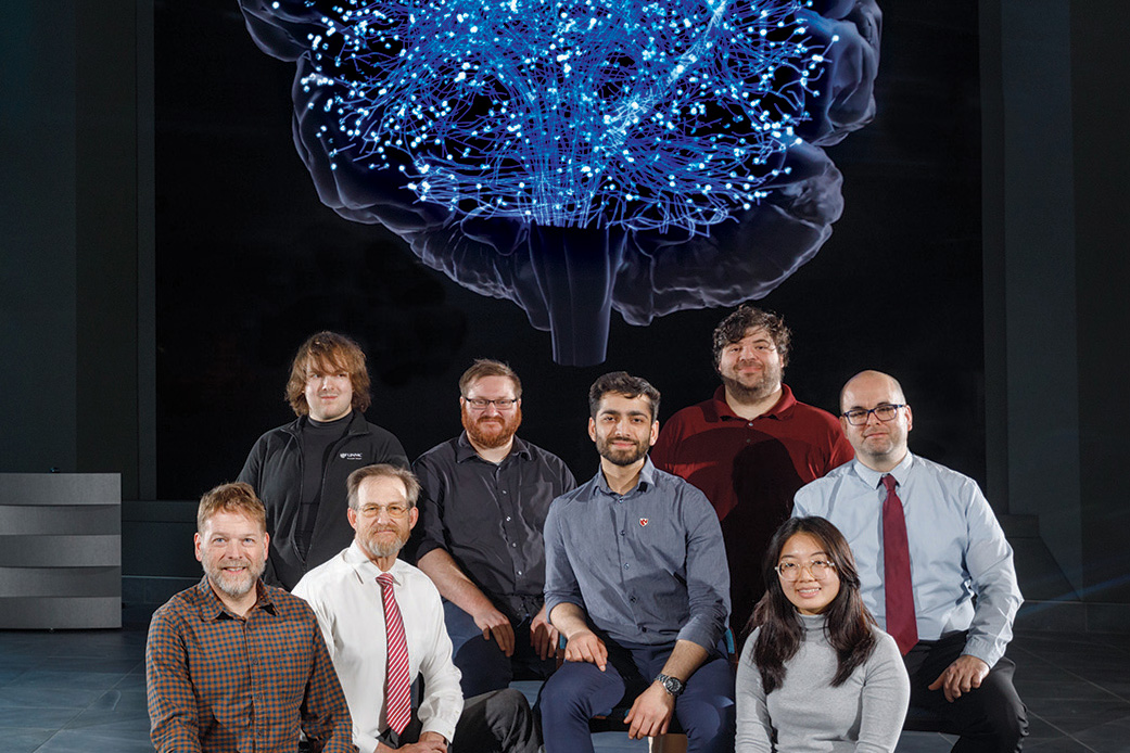
To craft this high tech content for UNMC students, iEXCEL calls on its team of designers, artists and animators with experiences from Hollywood to Netflix to the gaming industry to bring that science to life. Read on to learn about these Magicians of Medical Imagery.
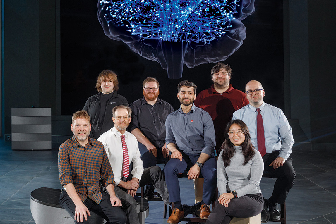
These Magicians of Medical Imagery - the iEXCEL Visualization Team - are (from left) Brian Curtis, Bill Glass, Wesley Fisher, Anthony Lanza, Paul Dye, Jeremiah Wilt, Jer Weann Ang and Dheeraj Varandani.
As a child, Bill Glass spent hours paging through Peterson Field Guides, the classic series known for its intricate wildlife illustrations. Glass remembers being captivated by the detail in drawings of such striking North American birds as the bald eagle and red-tailed hawk.
“It was like telling a story without words,” he said. These illustrative books would lead Glass, who “hated” math and English and “didn’t care” about history, to a career in medical illustration.
“All I liked was art and science.”
• • •
Growing up in Malaysia, Jer Weann Ang, too, was fascinated by the amount of information gleaned dissecting microscopy images and diagrams in biology books. “I wanted to replicate that and share the beauty of biology and life with others.”
Plans for a genetics and molecular biology graduate degree changed when an undergraduate art adviser in the U.S. suggested another way to combine her love of science and art: medical illustration.
Today, Glass and Ang transform complex biological and medical concepts into more easily understood 3D modules to benefit UNMC health profession students, faculty and staff — a stark contrast to hand-drawn anatomy illustrations studied by past generations.
This video is a conversion of the iEXCEL demo reel shown in the holographic theater at UNMC's Davis Global Center.
Their talents contribute to iEXCEL’s visualization team — a cadre of designers, artists and animators who create custom, anatomically correct, 3D-content for health education classes. Beyond medical illustration, the team’s resume boasts a range of skills and experiences that stretch from Hollywood to Netflix to the gaming industry — skills they use to bring science to life for UNMC students.
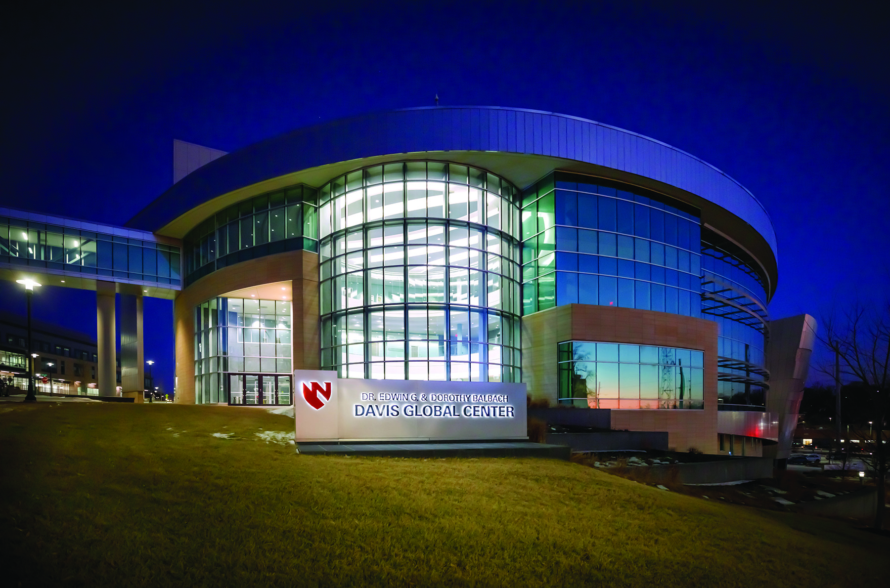
At UNMC — inside the Dr. Edwin G. & Dorothy Balbach Davis Global Center — the team is grounded by a clear mission and meaningful work. “We’re focused on making assets as accurate and informative as possible,” Glass said. “Seeing people use our content and having that ‘Aha!’ moment is rewarding."
They’re also on the front lines in charting a new course for health science education.
“iEXCEL set out to create the most accurate medical imagery possible because we believe 3D and virtual and augmented reality can help students learn about complex anatomy and physiology in an engaging way,” said Pam Boyers, PhD, associate vice chancellor of clinical simulation and iEXCEL. “We have combined gaming, modeling, simulation and visualization to create volumetric 3D imagery that brings learning to life.”
Students can — at their own pace and in their own time — manipulate and engage with organs and systems to better understand how they function. Real, but anonymized, patient data helps clinicians with diagnostics and pre-surgical planning.
“Ultimately, our vision is to distribute content across distance in real-time to respond to emergencies and provide tele-mentoring and tele-proctoring to save lives,” Dr. Boyers said. Success, she said, will depend, in part, on talented artists and technologists.
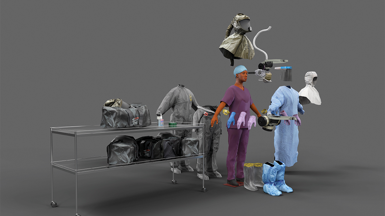
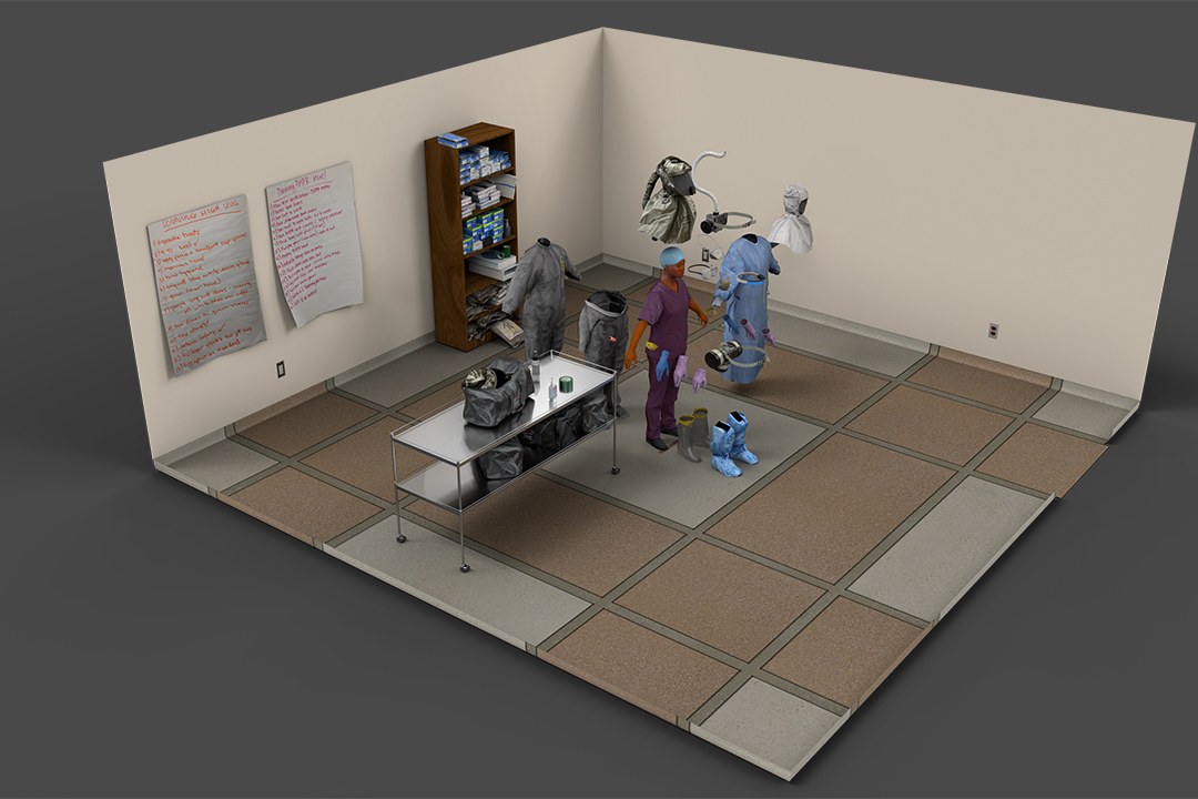
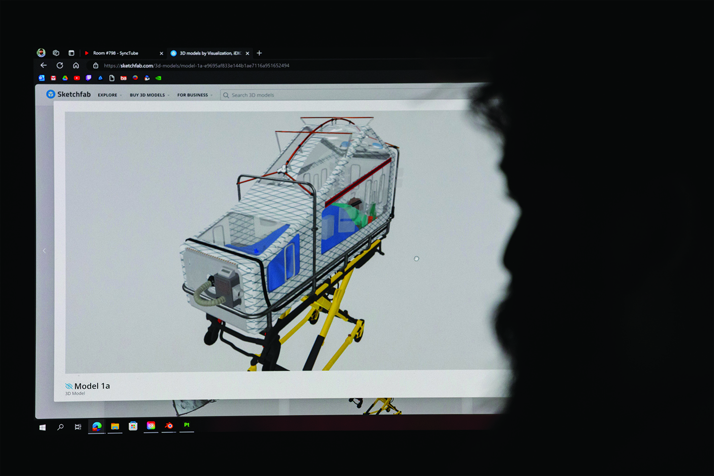
Using this interactive 3D program (see above and below), students and health care professionals can practice donning and doffing procedures for personal protective equipment.
Medical illustration has long played an invaluable role in translating science to improve lives. From illustrations on papyrus to Leonardo da Vinci’s anatomical figures to Andreas Vesalius’ anatomy book, “De Corpus Fabrica Humani,” to today’s 3D modules, illustrations transform complex information into visual images that improve learning and understanding.
German artist Max Brödel is considered the father of modern medical illustration. Hired at Johns Hopkins Hospital in 1894, he became the inaugural director of the first medical illustration program in the country in 1911. Many of his students became founding members of the Association of Medical Illustrators in 1945.
Glass wasn’t aware of such history while reading Peterson Field Guides. A love of oil painting and watercolors led him to a fine arts degree at Auburn University, and, from there, an understanding of the starving artist stereotype. After illustrating a pamphlet for a cousin’s home health company, he enrolled in the Medical College of Georgia, where he graduated with a Master of Science in medical illustration in 1989.
“I have been in bliss ever since,” he said.
Turns out medical illustration also was a perfect fit for Ang, who earned an undergraduate degree from the University of Wisconsin-Madison in genetics, with a minor in studio art.
She joined UNMC after graduating with a Master of Science in biomedical visualization from the University of Illinois at Chicago, in 2021.
“It’s a cool career because it’s so niche, and you can combine art and science,” Ang said.
• • •
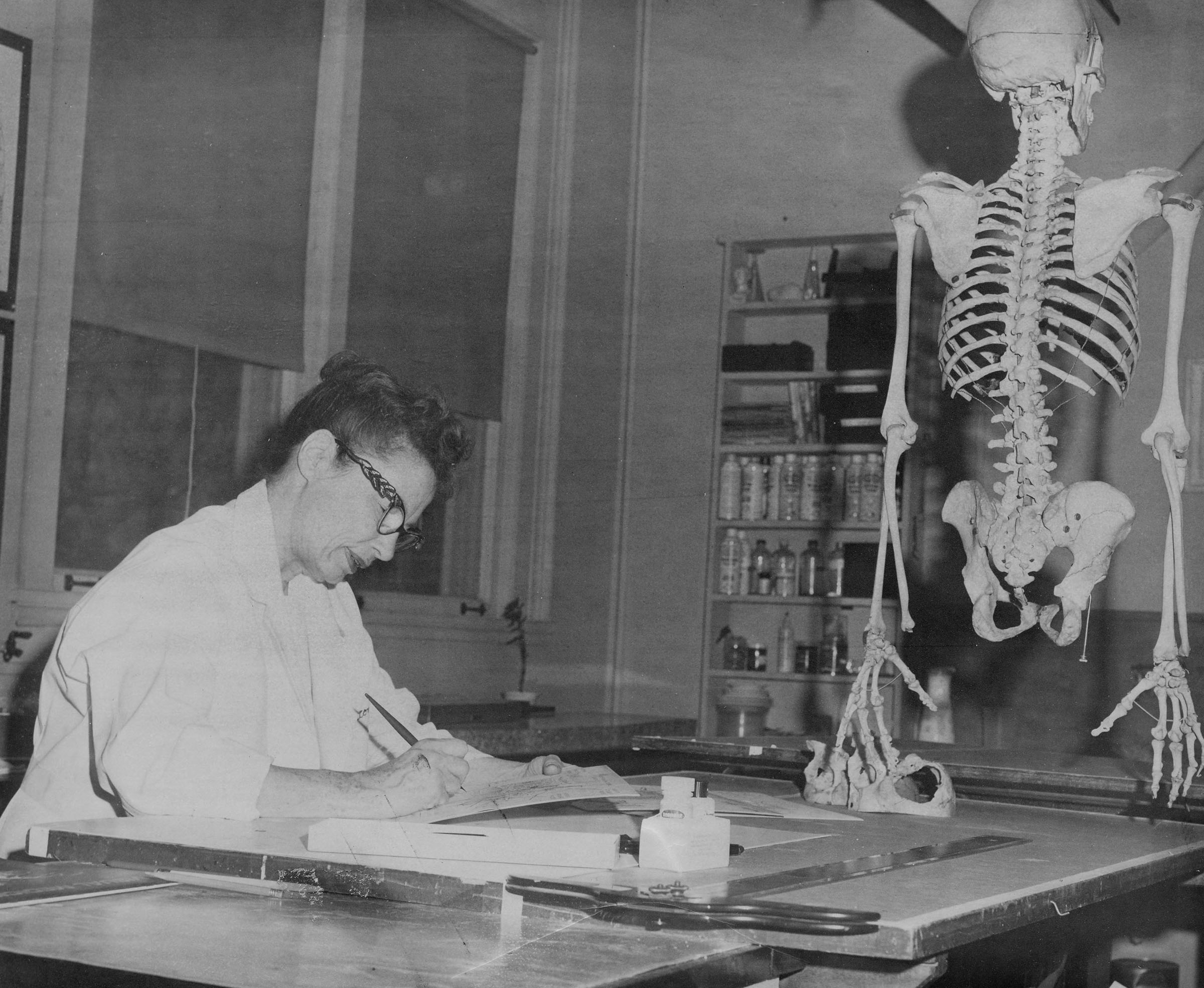
UNMC’s roots in medical illustration
Rose Reynolds spent 50 years (1929-1977) sharing her artistic skills with the UNMC community. An anatomical illustrator, she was one of the founding members of the Association of Medical Illustrators, who came together in 1945 to promote the “advancement of medical illustration and allied fields of visual education.” Years later, Bill Wassom (1984- 2013) provided medical illustration services to the UNMC community, initially through biomedical communications and later as part of UNMC Printing Services.
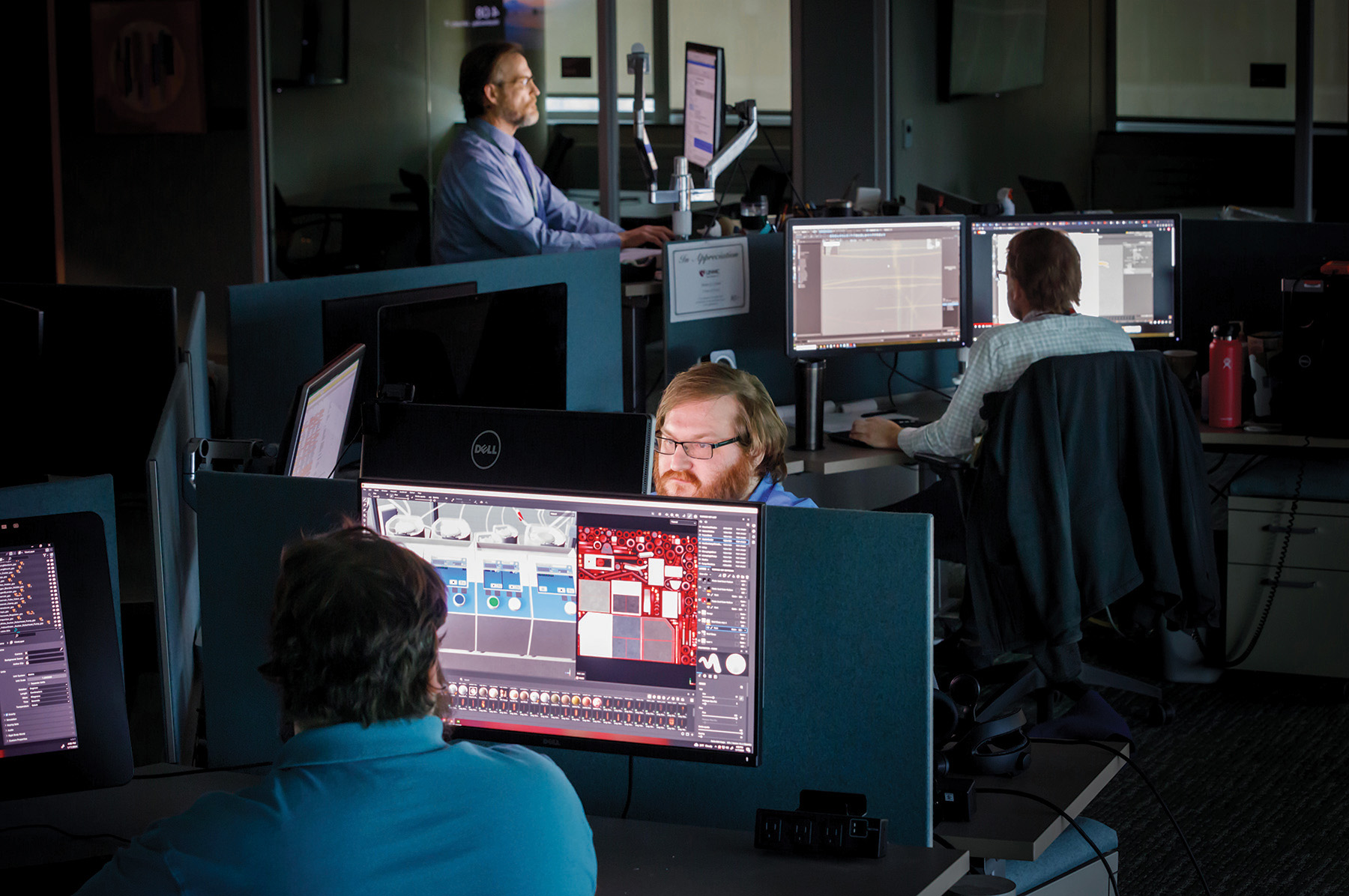
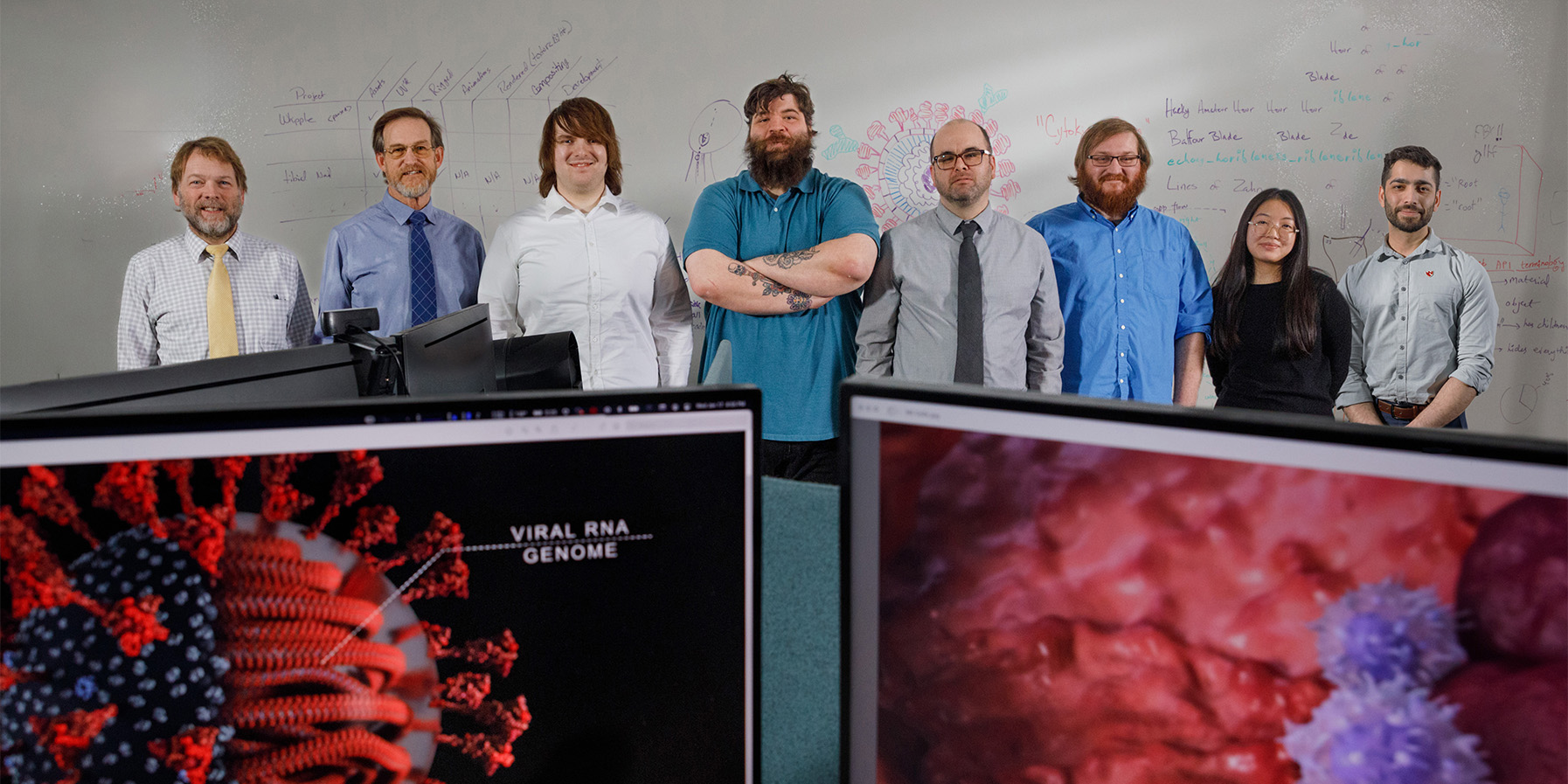
On this day, the lights are dimmed within UNMC’s Content Incubation Lab, and the room is quiet, but for the clicking of computer keyboards.
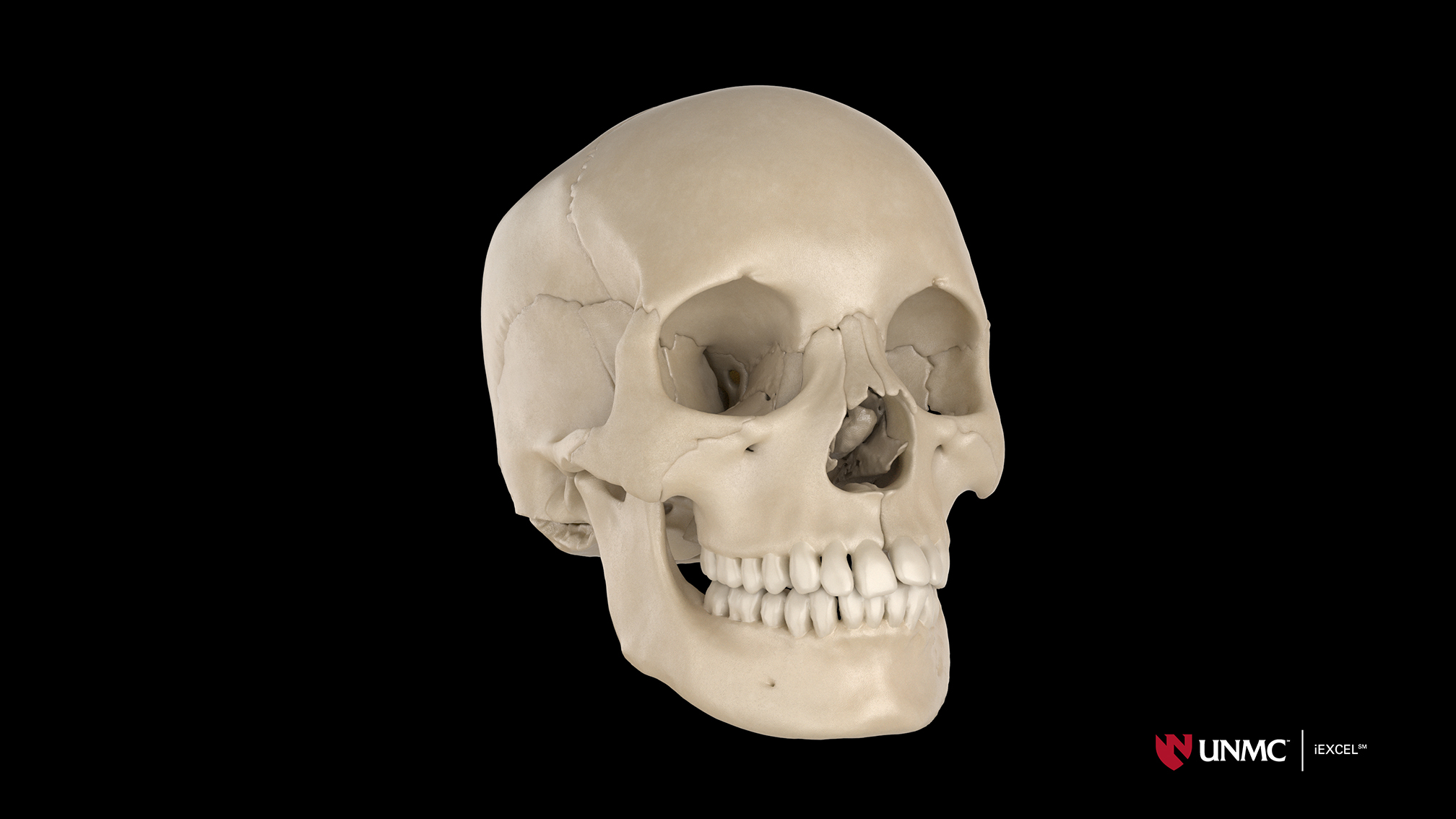
Upon closer look you see Glass, Ang and the rest of the creative team, busily at work — illuminated by the glow of their computer monitors. On one screen, you see an artist’s topographic evolution of a pelvic muscle. Nearby, data helps create a 3D skull. Steps away, a 3D perfusion machine springs to life onscreen with animation and coding that allows learners to turn knobs and “operate” the machine. Steps away, a vibrant blue eyeball is rendered as realistic as the eyes of its creators.
• • •
Merging art, science and technology is a painstaking process, and the iEXCEL team routinely works with subject matter experts to build accurate medical content, which includes building a near complete virtual 3D body — one deidentified module at a time.
With each proposed project, the team evaluates its purpose, audience and impact. Can it be used across multiple disciplines? Across multiple platforms? Does it grow iEXCEL’s anatomical catalogue? Can outcomes be measured? Does the bandwidth exist to create it?
Soon, the room’s ceiling-to-floor marker board begins to resemble an anatomical textbook with sketches, notes and storyboards that help subject matter experts and artists speak the same language.
From there, and at each stage of development, different team members add salient details that advance or refine the project. The collective work enables learners to ultimately see virtual 3D anatomical modules in exquisite detail. And, with each project, iEXCEL’s digital library grows, providing learners with new ways to study the heart, blood cells, eye, pelvis and more.
The team also creates digital twins of medical equipment and technologies to help students learn in safe environments, develop their confidence, practice competencies and be more prepared to enter the real world of health care.
“Every day is different,” Glass said, noting the team’s collaborations extend to other disciplines and industries as well as the military. “There’s not a recipe for doing VR applications or other experiential things so we do a lot of problem-solving, trial and error and learning of new software.”
Once a module is completed, the high-definition images are used throughout UNMC classrooms, and on computers, digital headsets, 3D CADWalls, iWalls and UNMC’s first-of-its-kind, four-story SONY wall.
Already, the team has:
- Created 3D animations that enable first-year students to study blood clotting.
- Created a unique interactive learning module to educate learners on benign paroxysmal positional vertigo, a leading cause of dizziness in adults.
- Developed a virtual crash cart that allows learners to explore before an emergency.
- Created anatomical models — an eye, inner ear, brain and skull — that can be dissected, labeled and manipulated in 3D space.
More than 50% of the team’s content runs on multiple platforms, Glass said.
• • •
Visual content brings science to life for UNMC medical student Uyen Tran.
Visual resources are a game-changer for UNMC’s Uyen Tran, a second-year medical student. “I’m a huge visual learner, and the iEXCEL team takes difficult concepts and makes amazing visualizations that really help students learn.”
A narrated coagulation video is among the standouts, she said, describing how it breaks down the complex sub-cellular process. “The visualization team, they’re perfectionists … and this showcases the factors coming in sequentially, one by one, and how they cleave to form a clot.”
For students, that detail and imagery create deeper understandings — especially with difficult-to-grasp concepts, she said. “When you read words off a textbook, you’re memorizing information because you can’t really visualize it, but with visualization you conceptualize it and understand why and how things happen.”
Knowing such material was available influenced the Hastings, Nebraska, native and University of Nebraska at Omaha graduate on where to pursue her interest in surgery. “I came thinking that would be really helpful for my education in medical school and beyond.”
She still remembers the day she and her classmates first saw an iEXCEL e-module. “Students were like, ‘Whoa! We made this? How do we even make this?’
"It was cool to see students be amazed at what our institution can do.”
Fantastic team and incredible work. We are fortunate to have this resource in our community!