 |
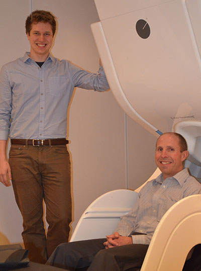
Standing is Alex Wiesman, a graduate research assistant in the lab of Tony Wilson, Ph.D., (sitting) director of the Center for Magnetoencephalography (MEG) and an associate professor of neurological sciences at UNMC. |
The study conducted over four years shows differences in brain activity in the occipital cortices, the area known for visual function, in HIV-infected patients who do and do not have HAND (HIV-associated neurocognitive disorders) as compared to healthy adults.
The findings, which are currently available online, are set to be published in the June issue of the journal “Brain.”
“Understanding the neural bases of these deficits could lead to earlier and more accurate diagnoses, more precise prognoses, as well as a more comprehensive understanding of the disease,” said Alex Wiesman, a graduate research assistant in the lab of Tony Wilson, Ph.D., director of the Center for Magnetoencephalography (MEG) and an associate professor of neurological sciences at UNMC.
While combination antiretroviral therapies have revolutionized the treatment of HIV infection, giving patients much longer lifespans, cognitive impairment remains a significant concern.
HAND traditionally has been diagnosed using a battery of neuropsychological tests and other measures and is comprised of three subtypes based on severity. It often includes notable deficits in attention, working memory, motor function and visual perception.
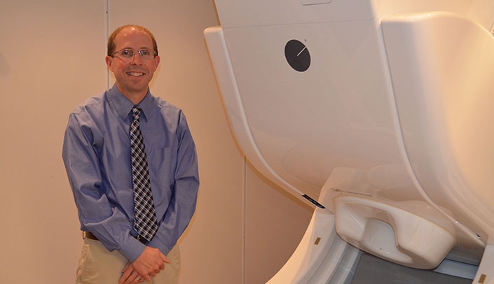
“The findings are very exciting and open up a lot of new possibilities in this area,” Dr. Wilson said. “MEG systems are becoming more and more common and using these systems to identify the presence of disease or track progression in neuro-HIV and other conditions is definitely on the horizon.”
By identifying a key neural response in visual brain regions that distinguished HIV-infected adults based on their HAND status, this finding may function as a diagnostic marker for HAND, Wiesman said.
“HAND currently affects more than 30 percent of all HIV-infected patients, thus an objective marker of the disease could have major diagnostic and prognostic implications,” he said.
MEG is a relatively new noninvasive brain imaging method that is silent and provides precise information about brain function by measuring ultra-minute magnetic fields that are generated by the brain. UNMC has been a national leader in MEG research for the past several years, Dr. Wilson said.
“Publishing in ‘Brain’ is a huge honor for our team and UNMC as a whole,” he said. “The fact that a graduate student lead-authored the paper also speaks to the quality of our graduate program at UNMC and the opportunities that are available to them.”
