When it comes to finding new treatments and cures for terrible diseases like cancer, one doesn’t want to waste time.
Now there’s a new, sophisticated instrument that may save some researchers a lot of time and effort.
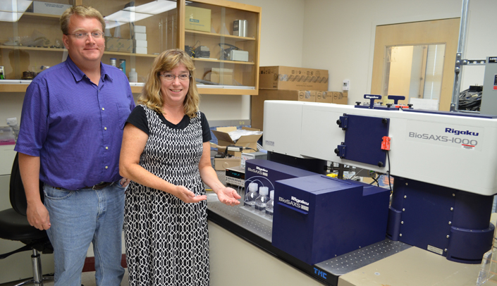
Available to researchers at UNMC and others, the BioSAXS 1000 is one of less than 10 in the world. It can analyze and provide a low-resolution 3D image that enables a researcher to understand the structure and function of proteins they are studying.
It also can clarify a researcher’s lab results to proceed to the next step of their research.
UNMC professor and researcher Gloria Borgstahl, Ph.D., procured the instrument, which sits aside the crystallography instrument in the Structural Biology Facility.
Jeff Lovelace, research associate in the Eppley Institute for Research in Cancer and Allied Diseases, said the equipment upgrade, which cost $535,000 is a great resource for UNMC researchers and others in the region.
He said the instrument can be used to determine the shape of macromolecules in solution without the need to first create a crystal.
“There are a lot of proteins we work with that won’t crystalize so we can’t use the crystallography instrument but we can use the BioSAXS on nearly all of our samples,” Lovelace said. “It gives researchers a better picture of what they’re looking at.
“It’s a valuable tool for help in understanding the shape of the biological macromolecule, seeing conformational changes, observing complexes, and designing drugs/drug delivery systems. The SAXS data can help us learn how to grow a crystal for high resolution studies at the atomic level.”
The instrument works by shining an X-ray beam on the sample and recording the scattering with an electronic detector. Data analysis includes calculation of dimensions of the molecule (radius), observations of problems with protein folding, and 3D modeling that displays the overall shape of the protein or complex.
It can be used to scan drugs to study their binding to the macromolecule, how they bind and if they disrupt a signaling complex. Promising drugs could then be tested on cells and mouse models.
“This instrument can help a researcher understand how their compounds interact with the biological target so that they will know if they’re going down the right path,” Lovelace said. “Sometimes research experiments don’t always work but they should if you’re on the right path. The machine can check for that. It’s a more targeted application versus a shotgun approach.”
