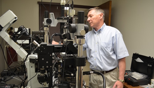
Before Yuri Lyubchenko, Ph.D., professor of pharmaceutical science in the College of Pharmacy, explains his research, he has a question: “Are you familiar with the concept of a record player?”
When the answer is “Yes,” Dr. Lyubchenko is relieved. So many students these days know only iPods, MP3s and streaming. They’ve never put a needle on vinyl.
But that — a record-player needle — is the principle behind the atomic force microscope (AFM).
In much the way a phonograph needle can decipher music in a record’s grooves, the atomically sharp AFM is capable of visualizing molecules and, under the right circumstances, even atoms. It has the capability to watch the interaction of molecules by performing time-lapse observations in water.
Among its nine various AFM instruments, UNMC houses a unique atomic force microscope, high-speed AFM. This instrument is capable of the time-lapse nanoscale imaging of molecules with video rate. It is one of the foremost tools for imaging, measuring and manipulating matter at the nanoscale.
Until the University of California, Berkley recently acquired a similar instrument, UNMC’s was the only one of its kind in the U.S.
Yet it’s kept without fanfare at the College of Pharmacy, behind an ordinary (locked) door, housed in a closet-sized room. Chances are you haven’t even heard it’s on campus.
But it allows Dr. Lyubchenko’s lab and others to do the kind of work that moves the needle — so to speak.
How so? Scientists know the DNA-binding enzyme APOBEC3G is a natural anti-HIV defense. But with the AFM, Dr. Lyubchenko and his team study the nanoscale structure and dynamics of APOBEC3G complexes with their DNA targets — the first step toward developing AIDS treatments based on nature.
AFM is also able to uniquely manipulate single molecules and measure their interaction.
This is especially important when looking into neurodegenerative disorders like Alzheimer’s, Huntington’s and Parkinson’s diseases.
Those diseases are caused when proteins, called amyloids, fold abnormally and aggregate. Knowledge of these “misfolded” molecular structures, and understanding how they assemble, is key to developing treatments, preventions and diagnostics of these devastating diseases.
UNMC scientists have pioneered an AFM force spectroscopy approach to probe misfolded states of amyloids, and to characterize their properties when they are first starting to come together as dimers (two-molecule complex). This is when these diseases first take root, and where treatments should be focused.
AFM, Dr. Lyubchenko said, is perfectly set up for testing drug-treatment candidates for this, and selecting the most efficient ones.
