Ramanathan Lab
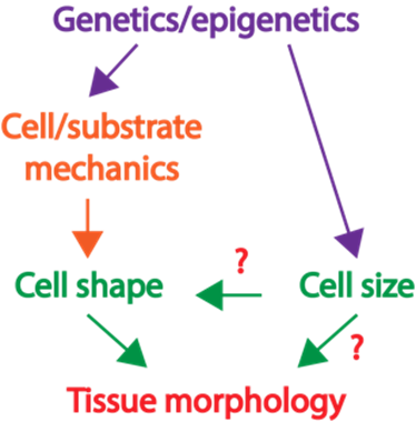
Fig. 1: The role of cell size in tissue packing is largely unknown.
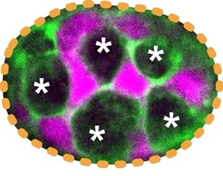
Fig. 2: The arrangement of the stem (magenta) and Paneth cells (*)cells at the base of an intestinal crypt
How animal cells of diverse shapes and sizes pack to form functional tissue is a fundamental biological question. Nevertheless, the current understanding of how tissues organize is mostly from examining the adhesive and tensile properties of similarly shaped cells. Previously, we discovered that discrepancies in cell size induce cell junctions to rearrange and cause stereotypical cellular reorganization. In our laboratory, we will dissect the principles of how, despite the rapid turnover, a checkerboard-like arrangement is maintained by the intestinal stem and support cells (Fig. 2).
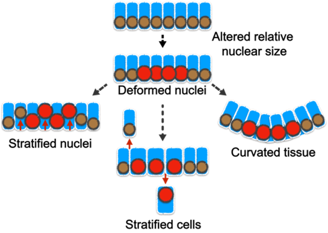
Fig. 3: The misregulation of cell/nuclear size and the hypothesized change in epithelial architecture.
Although cells of the same type have remarkably uniform morphology, tumor cells are typically pleomorphic and can exhibit abnormal variations in the size of cells and nuclei. Nevertheless, the influence of such variance in disease progression is not well understood. Furthermore, the tight correlation between the cell and nuclear size is frequently perturbed in diseases; but what are the pathological implications of this decoupling? Previously, we showed that abnormality in cellular size can have a profound impact on how cell pack and tissue organize. In our laboratory, we will determine how the organization of small intestinal villi in mice can be altered by abnormal cell and nuclear shape (Fig. 3).Our approach to addressing these questions is highly interdisciplinary. We integrate confocal imaging with biophysics and sophisticated genetic manipulation to quantitatively distinguish the influence of cell mechanics, shape, and size in tissue packing and homeostasis. We leverage a wide range of models including traditional 2D culture, 3D organoids, and mouse intestinal tissue. Over the long term, we anticipate that our study will extend the understanding of how cells self-assemble into tissues with stereotypical forms and reveal how functional tissue architecture is perturbed in disease.
Principal Investigator
Subu Ramanathan, PhD
Assistant Professor, Department of Genetics, Cell Biology, and Anatomy