Category: Pathology
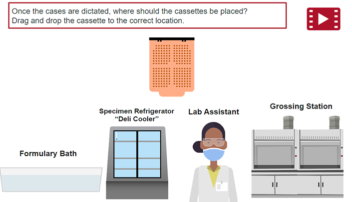
Advanced Practicum Course: Grossing Small Biopsies
This e-modules covers how to gross small biopsies and associated lab procedures.
Jul 19, 2022
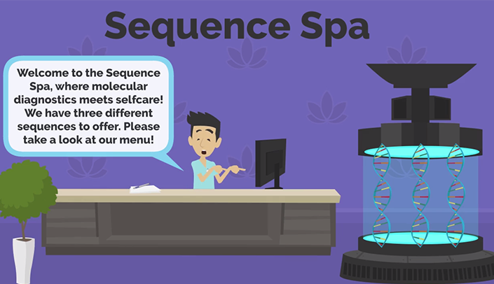
Companion Technologies: Molecular Diagnosis – Module 2
This e-module highlights the theory and analysis of Molecular Diagnostic techniques including- Pyro-Sequencing, Next Generation Sequencing, and informatics – within the environment of an animated medical spa.
Apr 12, 2021
Companion Technologies: Molecular Diagnosis – Module 1
This e-module introduces the theory and analysis of Molecular Diagnostic techniques including- molecular pathology, PCR, and Sanger Sequencing within the context of video games.
Apr 12, 2021

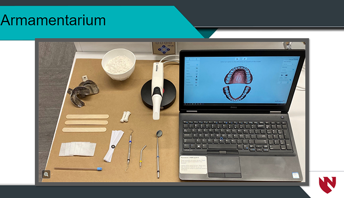
Digital Design – Occlusal Splint
Students can use chair side technologies to create a digital impression, or instead take a conventional impression/stone model to start the orthotic process. With an extraoral representation of the patients dentition, the data is transferred to a Computer Aided Design (CAD) software where the orthotic is designed. Once the design is completed, it will then […]
Apr 12, 2021
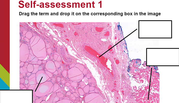
Cytology – Histology Morphologic Correlation of Thyroid Specimens
This E-module highlights the characteristics of the cytologic morphology and histologic morphology of thyroid specimens.
Mar 2, 2021
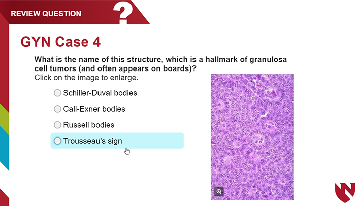
GU/GYN E-Module
In this E-module, 10 GYN cases and 7 GU cases are used to illustrate some of the different pathologies that may occur in the GYN/GU system. This module is designed for M2 students and Pathology residents.
Oct 1, 2020